PathoCaps.
The project of an MD pathologist, explaining Pathology to students, clinicians, lab professionals and patients alike.
“Learning together about our body, health and disease as a whole.”
Pathology, covered from every angle.
From surgical specimens to autopsy findings, PathoCaps walks through the subspecialties that make up the diagnostic backbone of modern medicine.
Diagnosis
Every post starts from a real diagnostic question — the kind a pathologist actually has to answer at the microscope, translated so anyone can follow the reasoning.
Evidence
Claims are grounded in current literature and established diagnostic criteria, not opinion. When something is uncertain, PathoCaps says so.
Objectivity
No sensationalism, no fear-based framing. Disease is explained as biology — precise, calm, and respectful of the patients behind every slide.
A trusted voice in Pathology.
Real numbers from a real, weekly-run account — not a media kit exaggeration.
 Gross specimen · from the archive
Run by an MD pathologist who stays behind the microscope.
PathoCaps is written and curated by a licensed Pathology specialist who keeps a private, low profile by design, in line with medical data-protection principles: the focus stays on the science and the cases, never on the person behind the microscope.
Since 2022, PathoCaps has grown into one of the most followed Pathology accounts in its field across Spain, the rest of Europe and the Americas — built one weekly post at a time, for an audience of physicians, residents, medical students, lab technicians and curious patients.
Where PathoCaps actually lives.
The full archive of cases, explainers and weekly stories is on Instagram. A live, embeddable profile widget isn't offered by Instagram to third-party sites anymore — so here's an honest snapshot, and a direct link to the real thing.
Interesting cases and medical information for everyone. Academic purposes only. Stories and cases, weekly.
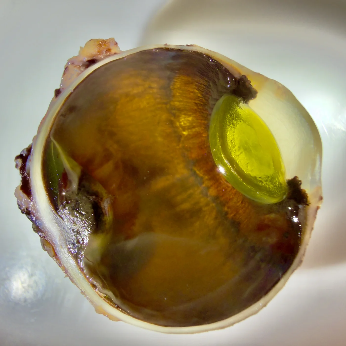
Gross dissection · the eye
A sectioned globe, opened to show the lens sitting in place — a favorite for teaching ocular anatomy.
View on Instagram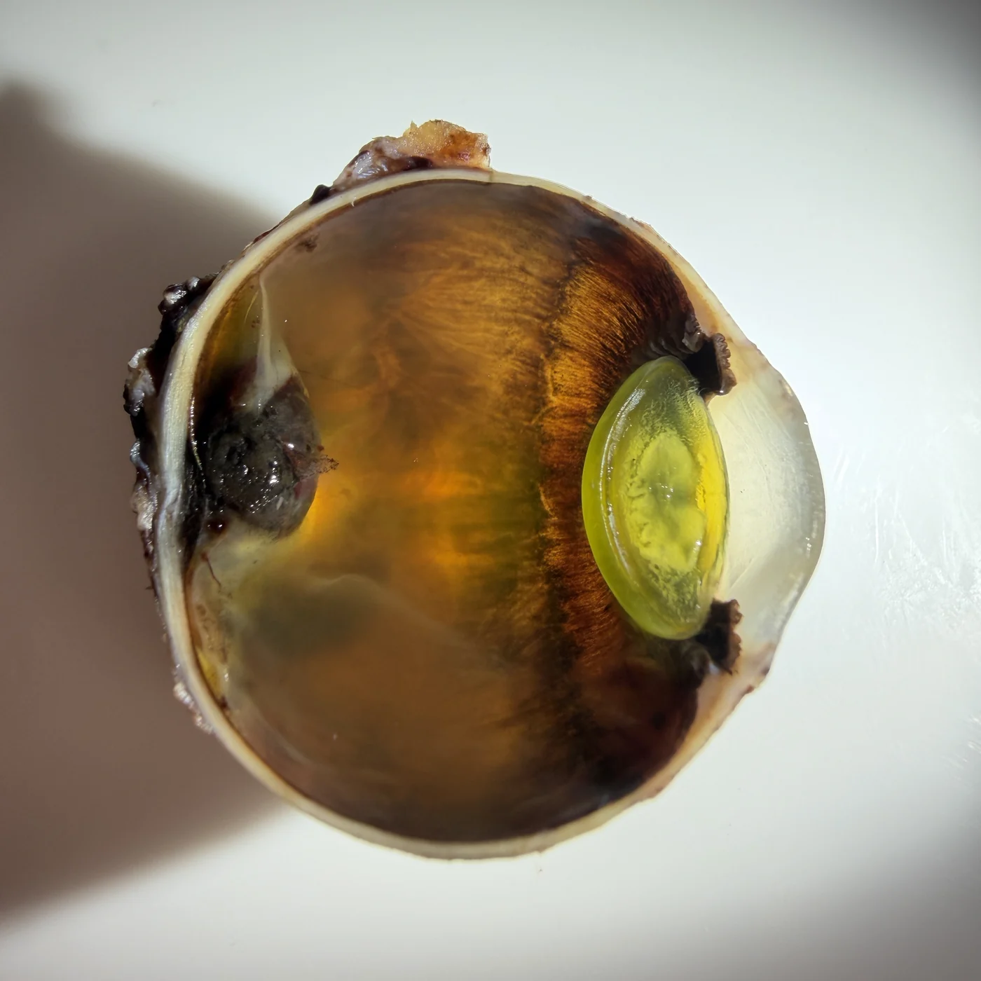
Same specimen, second look
A different angle on the same dissection — small changes in lighting reveal different structures.
View on Instagram
Special stain, close-up
A special stain brings out structures invisible on routine H&E — pattern recognition starts here.
View on Instagram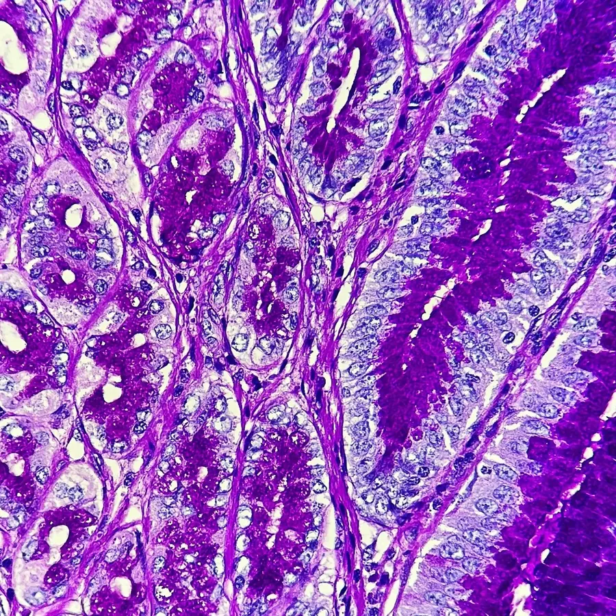
Routine stain, high magnification
Glandular architecture under higher power — the kind of field a report is built from.
View on Instagram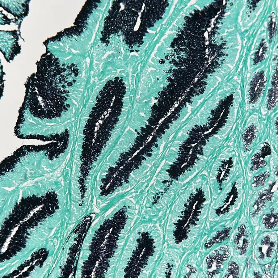
Zooming out
The same stain, wider field — context matters as much as the close-up.
View on Instagram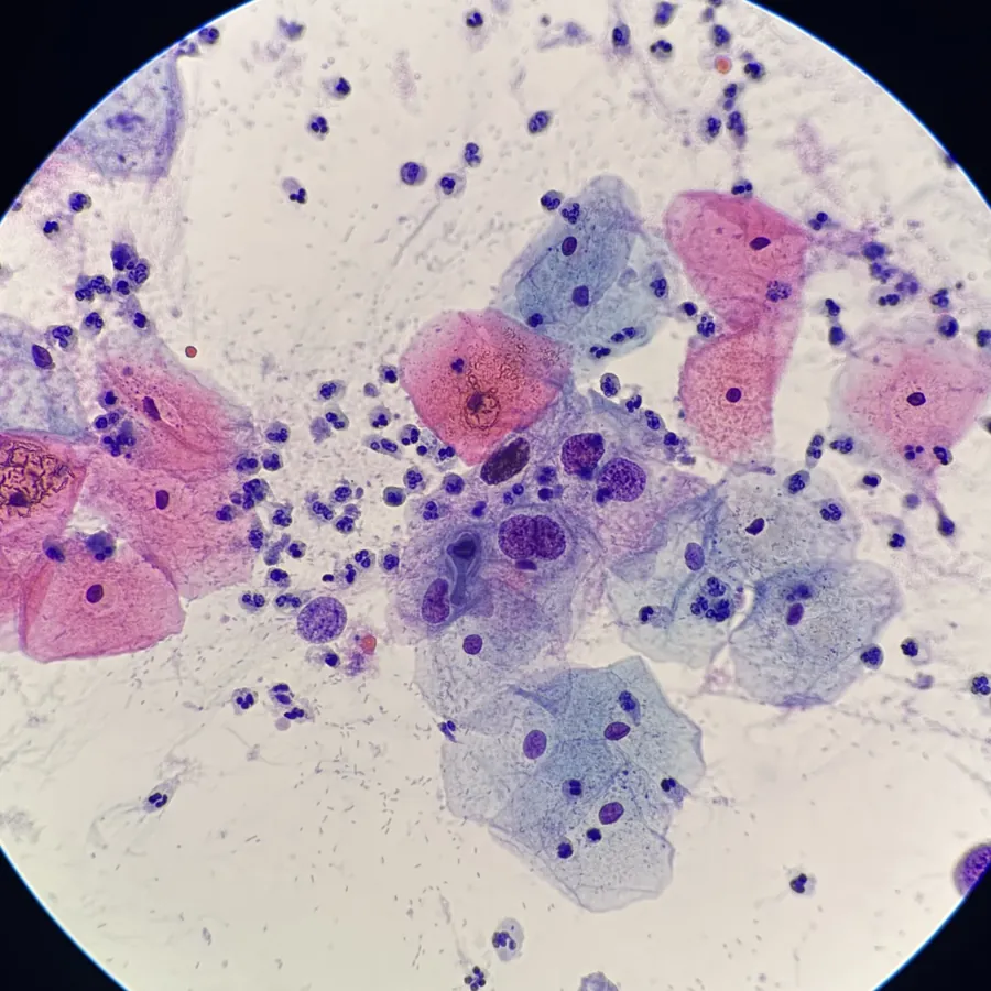
Cytology under the scope
A smear preparation — a different technique, the same habit of looking closely.
View on InstagramThese link out to the @pathocaps profile — new cases go up there first, so following is the only way to catch them as they happen.
“Let's learn Pathology together.”
Pathology decides more of modern medicine than most people realize — every biopsy, every surgical margin, every autopsy finding passes through it before a diagnosis becomes real. PathoCaps exists to open that black box: for the student staring at a slide for the first time, the lab technician who deserves to understand the why behind the protocol, the physician outside the specialty who wants a clearer picture, and the patient trying to make sense of a report written in a language that wasn't meant for them. No jargon left unexplained, no shortcuts on rigor — just an honest, weekly habit of looking closer.
— PathoCaps, est. 2022
Collaborations, press & questions.
PathoCaps keeps a low profile, but every message reaches a real person. Reach out for brand collaborations, academic partnerships or press.